
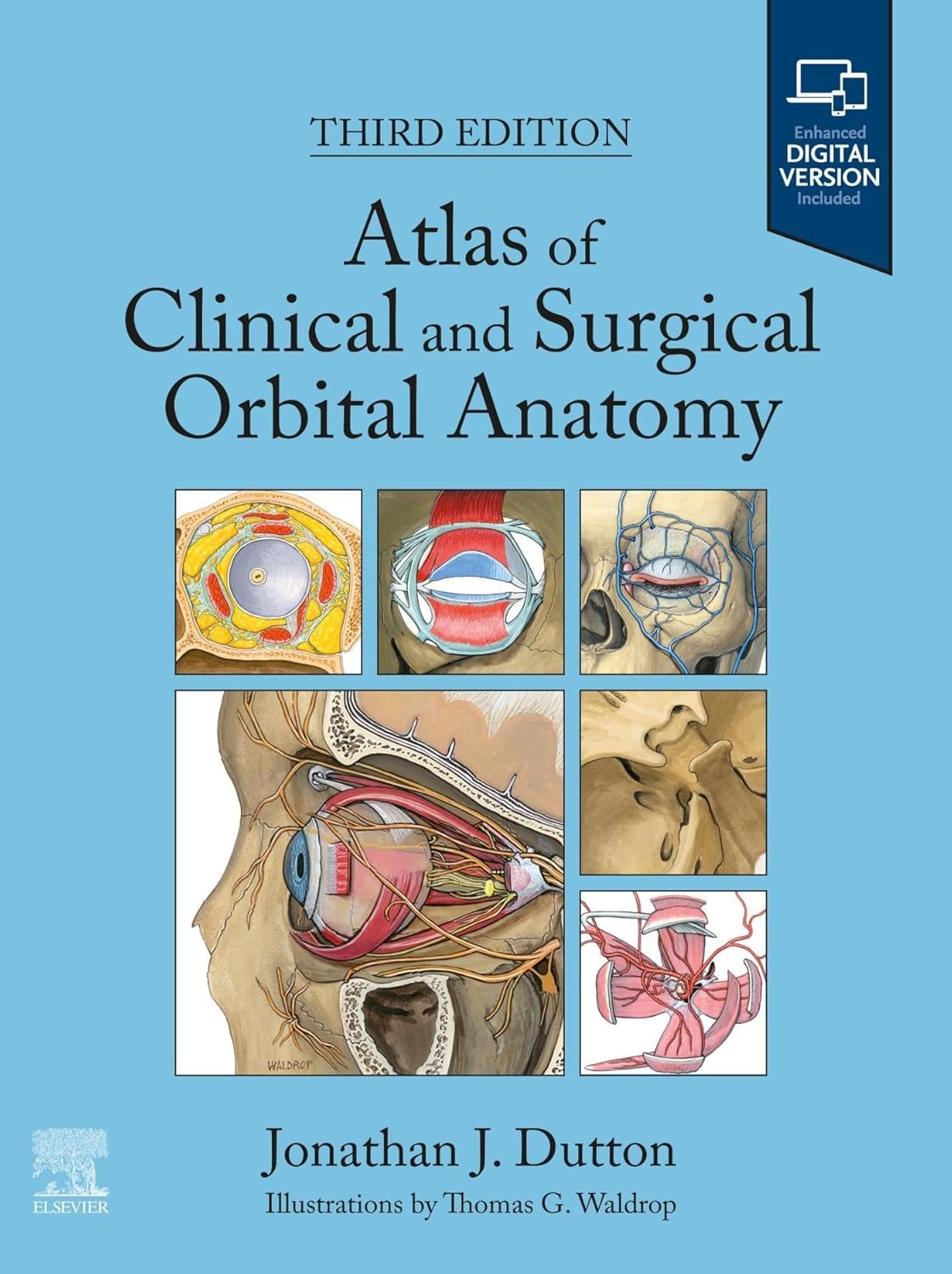
Enhance your surgical expertise with the 3rd edition of Atlas of Clinical and Surgical Orbital Anatomy, an essential resource for ophthalmic, oculoplastic, and other surgeons. This comprehensive guide offers a deep dive into orbital anatomy, from embryology to adult anatomy, under the expert authorship of Dr. Jonathan J. Dutton.
Key features of this richly illustrated guide include:
– Unique layered anatomical illustrations: Understand complex structures with the help of multiple sequential artworks. These illustrations highlight key intricacies and provide anatomical correlations with CT and MRI scans.
– Detailed three-dimensional depictions: Get a comprehensive view of each system through illustrations drawn from 150-micron histologic sections of human orbits. These are presented in frontal, lateral, and superior views.
– Clinical and surgical correlations: Expand your knowledge of how individual structures correlate with the most common clinical disorders and diseases. This feature is particularly beneficial for ophthalmologists, otolaryngologists, and plastic surgeons.
– Expanded Clinical Correlations chapter: This edition includes more disease conditions relevant to your field of work.
– New chapter on the Nasal Cavity and Paranasal Sinuses: Learn about the anatomy of these structures and how they relate to orbital disease, trauma, and surgery.
– Updated surgical procedure discussions: Gain insights into various surgical procedures, including orbital decompression, orbital floor fracture repair, strabismus surgery, and more. Understand how these procedures relate to orbital anatomy.
– Most recent literature references: Stay updated with the latest research and discussions in every chapter.
This fully revised edition is not just an anatomy atlas, but is also a visual guide to enhance your diagnostic and surgical expertise. Whether you’re studying embryology, adult
Authors:
Jonathan J. Dutton (Author)
Edition:
3rd
Publication Date:
October 6, 2023
From the book:


Reviews
There are no reviews yet.