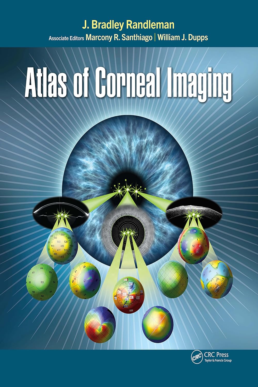
Introducing the Atlas of Corneal Imaging, a valuable resource tailored for physicians, surgeons, and trainees seeking in-depth knowledge on corneal imaging. This comprehensive guide, authored by esteemed experts Drs. J. Bradley Randleman, Marcony Santhiago, and William J. Dupps Jr, offers a thorough exploration of corneal imaging techniques, with over 1200 illustrative images and figures.
Unveiling a multitude of advanced diagnostic uses, this atlas navigates readers through the intricate process of analyzing corneal images using various technologies and devices. By showcasing a diverse array of devices and techniques, it provides a holistic view of the cornea’s structure, function, and pathology, enabling practitioners to identify subtle findings and evaluate signs of weakening or pathology effectively.
Key features of the Atlas of Corneal Imaging include:
– Interpretation of topographic patterns and mapping
– Evaluation of corneal ectasia and refractive surgery
– Correlations with corneal disorders
– Exploration of corneal surgery complications
– Comprehensive assessment for cataract surgery
This unique resource bridges the gap in corneal imaging literature by adopting an image-first approach, making it easier for readers to comprehend the different imaging technologies available. Whether you are a seasoned professional or a trainee, this atlas equips you with the knowledge needed to interpret corneal images accurately and make informed clinical decisions.
Explore the world of corneal imaging with confidence and precision with the Atlas of Corneal Imaging. Unlock the potential of cutting-edge imaging technologies to enhance your practice and improve patient outcomes.
Authors:
J. Bradley Randleman (Author)
Edition:
Publication Date:
June 1, 2024
From the book:









Reviews
There are no reviews yet.